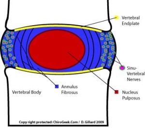
The annulus is a ligament and like any ligament in the body can be torn. Tearing of the annulus can produce pain because the annulus has pain fibers within its structure. Many episodes of low back pain are probably tears in the annulus. This condition is called an annular tear or internal disc disruption or sometimes degenerative disc disease.
This is what a normal disc looks like:
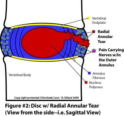
Most annular tears are caused by the natural aging process. Since the neck and back are responsible for bearing most of a person’s body weight, they are susceptible to a great deal of wear over time. By the age of 30, most people’s intervertebral discs have begun to degenerate to a certain degree. This degeneration can lead to annular tears since the annulus fibrosus is in a weakened state. A traumatic injury can also cause an annular tear. This is typically seen in those who participate in high-impact sports such as gymnastics and football and in people with strenuous occupations. Those experiencing annular tear symptoms should contact their primary care physician for information regarding the diagnosis and treatment of their spinal condition. Pain in annular tear is associated with sinuvertebral nerve endings inside each disc.
This is what a disc with annular tear looks like:
Diagnostics
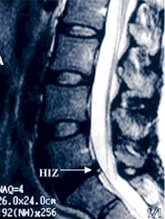
Annular tears are sometimes hard to diagnose! Modern imaging techniques and radiologists often fail to see an annular tear on MRI. Usually, contrast-enhanced MRI shows HIZ (high-intensity zone) which means that person or better say disc has an annular tear.
This is what it looks like:
However, a high-intensity zone on MRI does not mean that the disc itself is causing pain. That is why additional tests are to be done. The only method to conclude if the intervertebral disc is causing pain is called discogram or discography. This test will show if the disc is degenerated and reproduce patient’s everyday pains. If the test is concordant, this means that the disc with HIZ and discography confirmed concordant pains is indeed the cause of patient problems.
This is how discography confirms abnormal contrast distribution in the intervertebral disc:

Treatment
Now, the hardest part. How to tell the patient that the only treatment for such a condition is an aggressive approach and surgery…? Either fusion or artificial disc replacement surgery – both of them risky and invasive.
Thanks to the valuable work of Dr. Martin Knight, Dr. Daniel Choy, Dr. Peter Ascher, Dr. Thomas Hoogland, Dr. Sang-Ho Lee, Dr. Anthony Yeung, and, of course, our spinal neurosurgeon Dr. Robert Saftić who adopted their techniques, we are in a position to offer safe minimally invasive alternatives to spinal fusion and artificial disc replacement.
PLDD – laser disc decompression – in discography and MRI confirmed annular tear and internal disc disruption (in the early stages) we can proceed with fluoroscopically controlled minimally-invasive laser treatment with goals of shrinking the disc and ablating painful sinuvertebral nerve endings inside the disc.
SED – selective endoscopic discectomy – in discography and MRI confirmed annular tear and internal disc disruption (in more advanced stage) we can relieve your pains and symptoms with this endoscopically assisted and controlled minimally-invasive treatment which includes evocative discography to mark degenerated tissue inside the disc. Later on during the operation, a surgeon is able to identify degenerated tissue and annular tear, seal it and ablate painful nerve endings inside the disc.
At Aksis Special Hospital, spinal neurosurgeon Dr. Saftić successfully applies laser and endoscopic surgical techniques to treat annular tears. Do not hesitate to contact us for additional information and a free MRI review.