For the first time ever, invasive surgeries like microdiscectomy, laminotomy, foraminotomy, and inner decompression of the spinal cord can be performed in a less invasive way and without the use of general anesthesia. The METRx system combines the reliability of conventional microsurgical methods with the advantages of a minimally invasive technique. The advantage of METRX system over traditional microsurgical methods is that now the neurosurgeon can perform spine surgery by using segmental dilators and tubular retractors and apply a technique that minimizes the amount of damage to muscle tissue which is the main factor of spine stability. Contact us for more information.
Features and Benefits
With the METRx™ System, spine surgeons are now able to remove herniated or “slipped” discs in the lumbar spine with a minimally invasive technique that significantly decreases the amount of pain felt by the patient post-surgery. Over the last 10 years, many of the surgical sub-specialties in Orthopedics and Neurosurgery have used fiberoptic video cameras and specially designed tools to assist them in performing surgery with much smaller incisions, less damage to the surrounding tissue, and faster recovery times. Most people are familiar with this type of technology in the form of “arthroscopic” surgery, which allows orthopedic surgeons to look inside joints like the knee and operate through very small incisions with a minimal amount of pain, scarring, and trauma to the muscles that move the knee. This type of technology has recently been applied to spine surgery, and with the new METRx™ System created by Medtronic Sofamor Danek, surgeons can treat herniated discs in the lumber spine with much less trauma to the muscles that support and protect the spine. Minimally invasive surgical techniques have become the standard of care in the treatment of many conditions that affect the joints, heart, and GI tract. With this new technology, spine surgeons are now able provide their patients with the same benefits of less invasive surgery, less postoperative pain, shorter recovery times, and easier rehabilitation.
What type of Spine Surgery is the METRx™ System Used For?
The METRx™ system was designed in order to provide spine surgeons with a way of removing herniated intervertebral discs in the lumbar spine that are putting pressure on the spinal nerve roots and causing pain. Herniated discs occur because the discs that act as shock absorbers between the bones of the spinal column become stiff and less flexible as we grow older. Discs that lose their flexibility can rupture and cause neck pain and back pain. A part of the inside of the disc can herniate or leak out through a tear in the ligaments that surround the disc, pressing on the spinal cord or on the nerves that travel out to the arms and legs. A herniated disc can be responsible for a pain that is felt in the neck or back, but the pressure that the herniated disc puts on the spinal cord or the nerve roots can cause pain in the arms or legs and sometimes can even cause problems with the way the spinal cord normally functions. If a herniated disc is pinching a nerve in the neck, most people will have persistent neck pain, often with numbness in the arms and hands. If a herniated disc is pinching a nerve in the lower back, then most people will have persistent low back and buttock pain, with numbness and tingling in the legs and feet. When the symptoms of a herniated disc do not respond to physical therapy, medications, or improve with time, then surgery can be an option. This type of spine surgery is called a “discectomy” and it is used to remove the part of the herniated disc that is pinching or putting pressure on the nerve roots in order to relieve the pain and numbness. With traditional disc surgery, spine surgeons make an incision in the center of the back or neck at the same level as the herniated disc and then strip the muscles of the back away from the bones of the spinal column in order to be able to see the area where the disc has herniated. Once the herniated disc has been removed, the muscles are put back in place and the surgeon closes the incision.
How is surgery with the METRx™ System Different?
With the new METRx™ System, surgeons can perform this type of discectomy surgery with a special type of muscle-splitting technique that minimizes the amount of muscle damage that is necessary in order to be able to see where the herniated disc is located and safely remove the herniated fragments while protecting the nerve roots and spinal cord. In comparison to a standard discectomy, a microsurgery discectomy performed with the METRx™ System causes much less pain after the surgery, and allows patients to leave the hospital earlier, and return to work and their daily activities sooner. This type of surgery also makes rehabilitation and physical therapy after spine surgery much easier because there has been less damage done to the muscles that move and protect the spine, which leads to less scar tissue formation.
How does the METRx™ System Work?
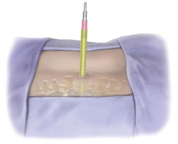
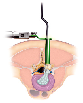
The main advantage of the METRx™ system in comparison to a tradition discectomy is a smaller incision and less damage to the muscles of the spinal column. This advantage is achieved by allowing the surgeon to expose the area where the herniated disc is located without making a large incision. A discectomy that is done with the METRx™ system begins with the surgeon precisely localizing the level of the herniated disc with a very small needle that is inserted through the muscles of the back down to the area where the disc fragments are located. The correct position of this needle is confirmed with a special type of X-ray machine that is used in an operating room called a fluoroscope. After the correct position of the needle has been confirmed with fluoroscopic guidance, a series of soft-tissue dilators are used to create a small tunnel that measures 16mm in diameter (less than ¾ of an inch) through the muscles of the back so that a hollow tube can be inserted down to the level of the spinal column. This tube, which is called a tubular retractor, contains a very special video camera with a magnifying lens and a fiberoptic light source that illuminates the tissues and relays the images to a separate video screen so that the surgeon can operate safely.